SURGICAL PHOTOS OF CASE NO 2
Fig 1: Pre-op Xray indicating Non-Union with bone loss
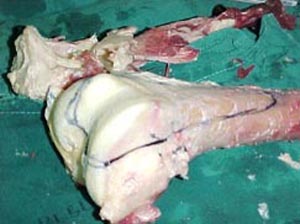
Fig 2: View of classical Knee allograft
Fig 3: Allograft being prepared
Fig 4: Resection margin on the allograft
Fig 5: Cutting to size of the allograft
Fig 6: Appropiate size allograft cut
Fig 7: Allograft being fixed with screws
Fig 8: Incision site being sutured
Fig 9: Post Operative X-ray image of Internal fixation of allograft
THANK YOU FOR BEING WITH US