SURGICAL PHOTOS OF CASE NO 1
Fig 1: Pre-Op X-ray indicating Osteosarcoma with pathological fracture
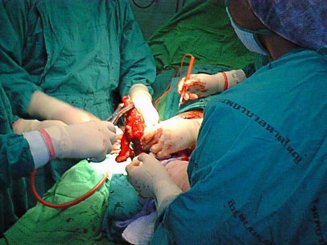
Fig 3: Prof Yongyudh and Dr. Phairot at work
Fig 6: Size determination of Lesion
Fig 7: Freeze dried whole humerus allograft
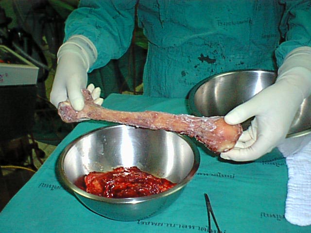
Fig 8: Soft tissue removed from the allograft
Fig 9: Allograft sized and soaked in Betadine
Fig 10: Reduction to check the size of allograft
Fig 11: Intramedullary remaing of the allograft
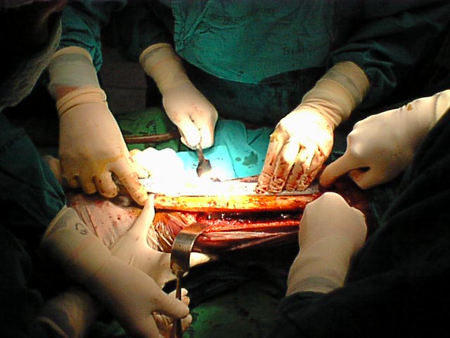
Fig 12: Allograft reduced and fixed with Intra-Medullary Interlocking Nail
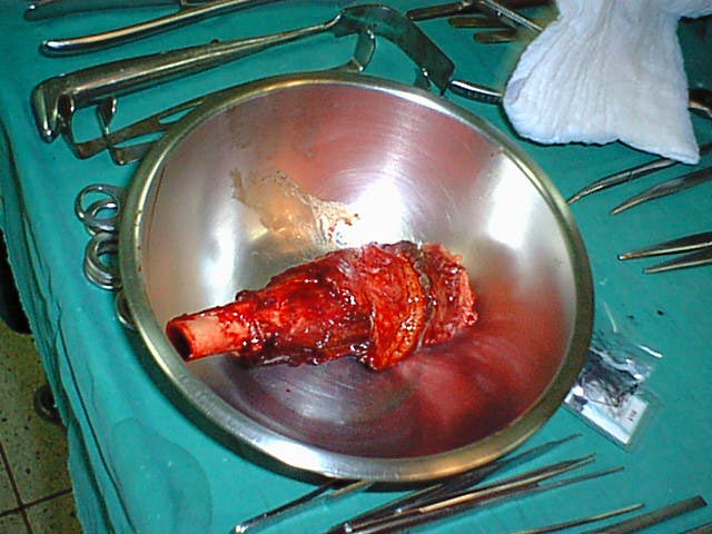
Fig 13: Transverse section of the resected lesion
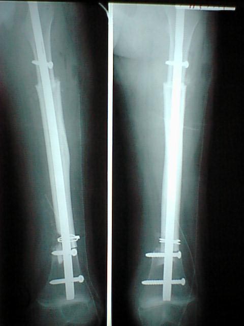
Fig 14: Post-operative x-ray showing solid stablization
THANK YOU FOR BEING WITH US